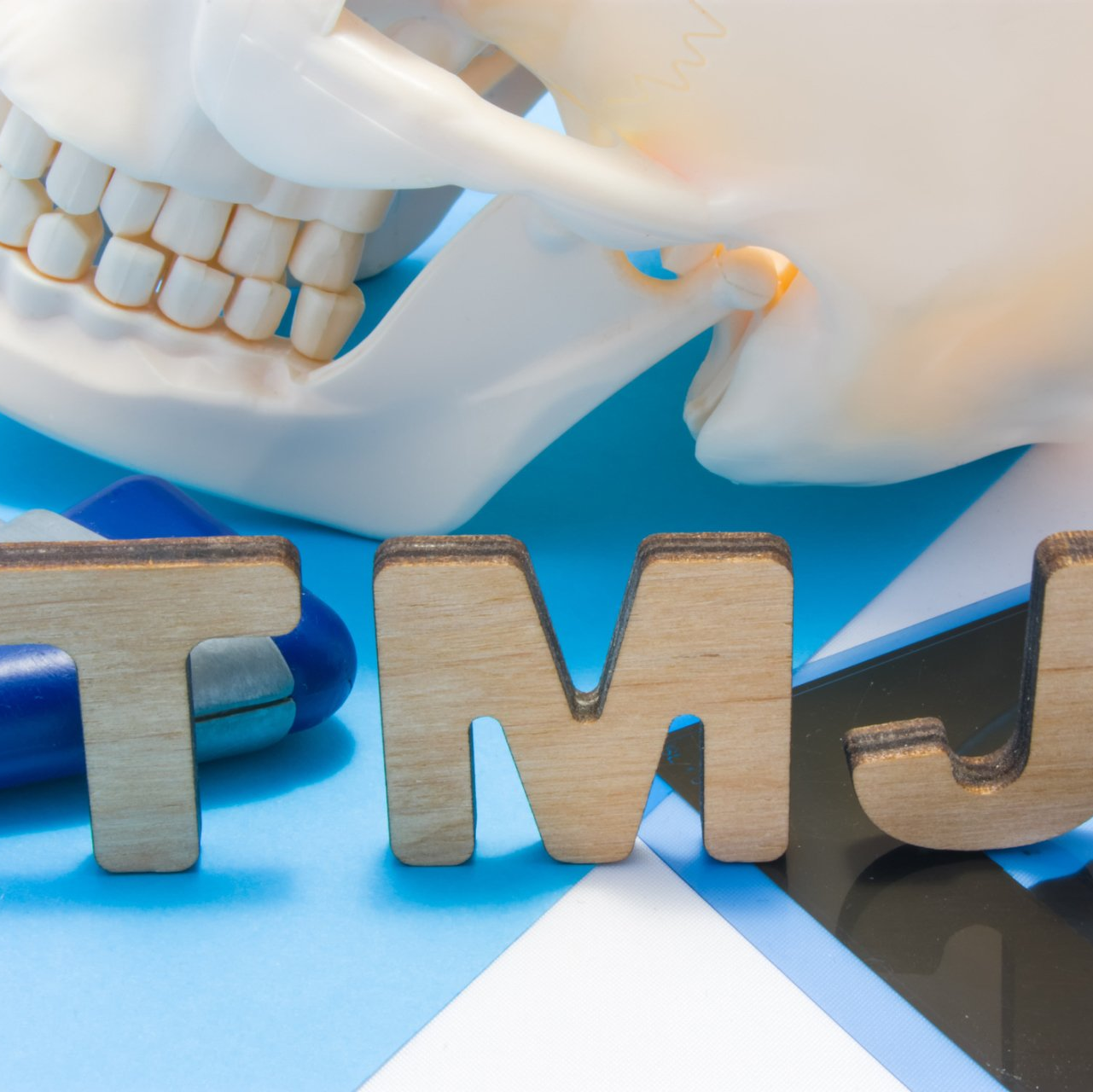
Oral Surgical Procedure Near North Raleigh, NC
Oral Pathology/Biopsy
The inside of the mouth is normally lined with a special type of skin (mucosa) that is smooth and coral pink in color. Any alteration in this appearance could be a warning sign of a pathological process. The most serious of these is oral cancer. The following is a list of changes that may represent the beginning of a pathologic process or cancerous growth:
- Reddish patches (erythoplasia)
- Whitish patches (leukoplakia)
- A sore that fails to heal and bleeds easily
- A lump or thickening on the skin lining inside of the mouth
- Chronic sore throat or hoarseness
- Difficulty chewing or swallowing
When an abnormality is discovered, a referral is often made to an oral and maxillofacial surgeon. Often, he or she will feel a biopsy is necessary for a complete and accurate diagnosis.
A biopsy is a surgical procedure that involves the removal of a piece of suspicious tissue; usually part of the lining tissue (mucosa) of the mouth or the underlying bone that has demonstrated possible involvement through the examination process. Fortunately, most biopsies can be carried out in the office setting with local anesthesia.
The harvested piece of tissue, or specimen, is sent to a pathology laboratory for examination where the tissue is handled by a qualified specialist in oral pathology. They will process the tissue and examine the specimen under a microscope. After the examination is complete, the pathologist will send a report to your surgeon for review.
The report helps establish a diagnosis as well as enabling your surgeon to develop a treatment plan that addresses the type of lesion that was identified. Small lesions may have been removed in their entirety during the biopsy while the larger lesions may have had only a small portion removed thereby necessitating further surgery.
Impacted Canines/Orthodontic Exposure
Exposure and Bracketing of an Impacted Tooth
An impacted tooth simply means that it is “stuck” and can not erupt into function. Normally, the maxillary cuspid teeth are the last of the “front” teeth to erupt into place. They usually come into place around age 13 and cause any space left between the upper front teeth to close tight together. If a cuspid tooth gets impacted, every effort is made to get it to erupt into its proper position in the dental arch.
In cases where the eye teeth will not erupt spontaneously, the orthodontist and oral surgeon work together to get these teeth to erupt. In a simple surgical procedure performed in the surgeon's office, the gum on top of the impacted tooth will be lifted up to expose the hidden tooth underneath. If there is a baby tooth present, it will be removed at the same time.
Once the tooth is exposed, the oral surgeon will bond an orthodontic bracket to the exposed tooth. The bracket will have a miniature gold chain attached to it. The oral surgeon will guide the chain back to the orthodontic arch wire where it will be temporarily attached. Sometimes the surgeon will leave the exposed impacted tooth completely uncovered by suturing the gum up high above the tooth or making a window in the gum covering the tooth. Most of the time, the gum will be returned to its original location and sutured back with only the chain remaining visible as it exits a small hole in the gum.
Shortly after surgery the patient will return to the orthodontist where a rubber band will be attached to the chain to put a light eruptive pulling force on the impacted tooth. This will begin the process of moving the tooth into it's proper place in the dental arch.
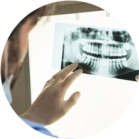
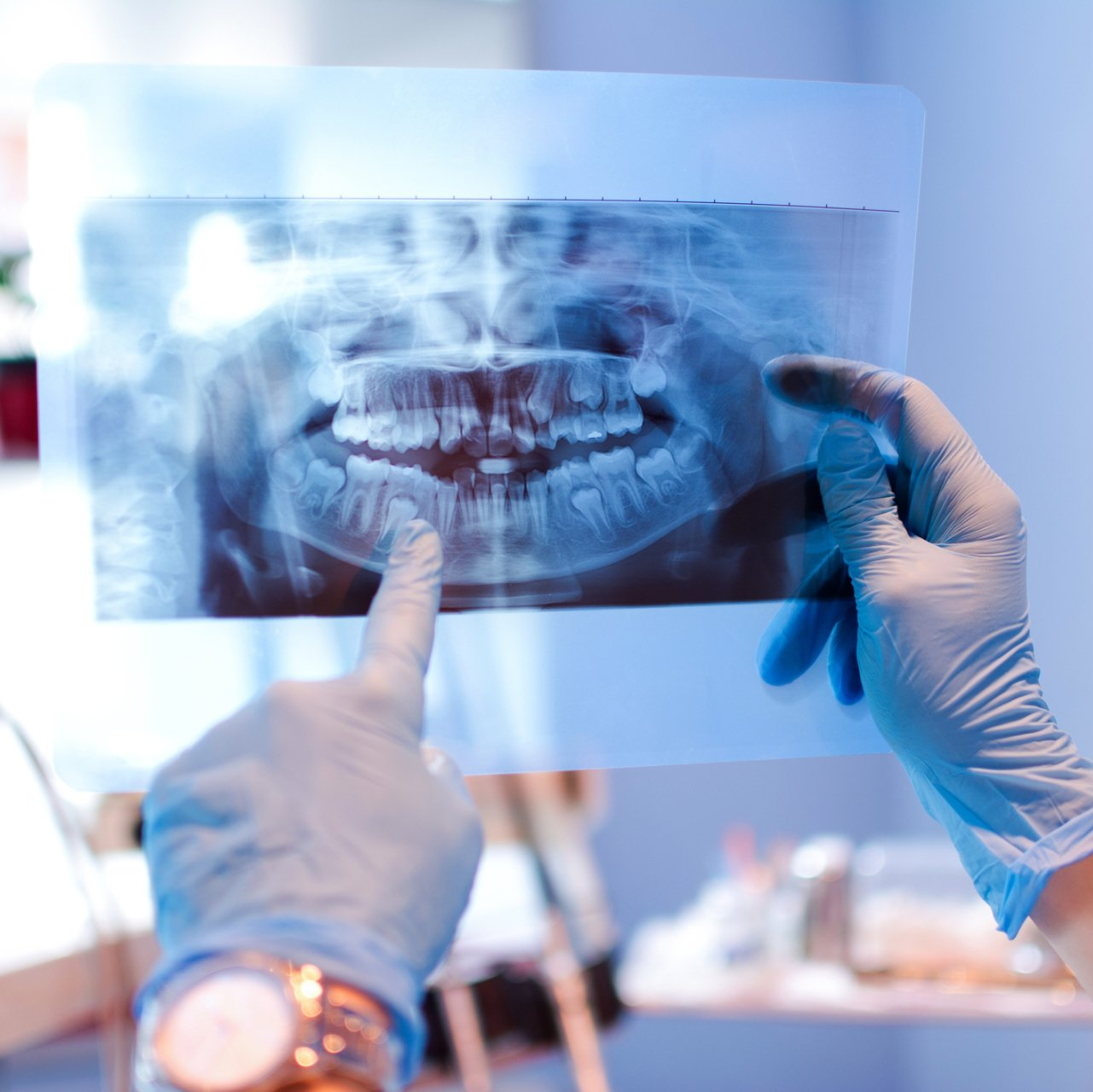
CT/Cone Beam Imagery
Cone beam computed tomography (commonly referred to by the acronym CBCT) is a medical imaging technique consisting of X-ray computed tomography where the X-rays are divergent, forming a cone. CBCT has become increasingly important in treatment planning and diagnosis in implant dentistry , among other things.
During a CBCT scan, the scanner rotates around the patient's head, obtaining up to nearly 600 distinct images. The scanning software collects the data and reconstructs it, producing what is termed a digital volume composed of three dimensional voxels of anatomical data that can then be manipulated and visualized with specialized software.